Magnetic Resonance Imaging (MRI):
What is a Magnetic Resonance Imaging (MRI) scan?
MRI is a non-invasive technique that uses a combination of magnetic fields and radiofrequency radiation to get images of soft-tissue in the human body. An advantage of MRI over other imaging techniques is that there is no ionizing radiation (radiation that can damage cells) used, so there are no exposure issues.
What is involved with the MRI procedure? Is there anything a person can or should do to prepare?
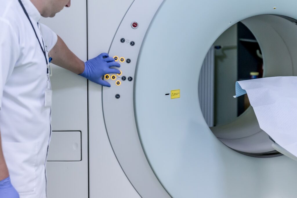
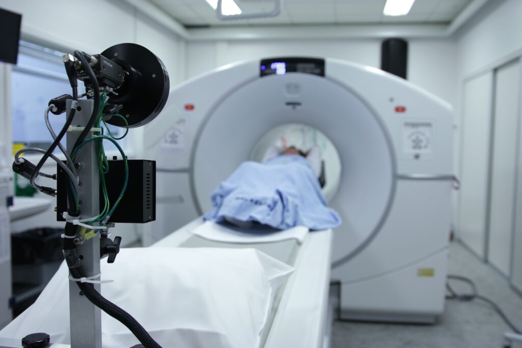
For an MRI of their brain, a person is asked to change into a hospital gown to ensure that no ferromagnetic materials (substances that have high potential to act as magnets) are introduced to the scanner while you are there, as that can be dangerous. The technical staff at the site can explain more about ferromagnetic materials and what should be done to minimize any risk. The person is then placed on a table that will move into a long tube. The procedure typically takes around 30 minutes and the machine will produce various noises as different types of images are obtained. No preparation is usually necessary, unless the person has problems with claustrophobia. In that case, individuals are encouraged to speak with the clinician or individual who has ordered the MRI to receive guidance on additional preparation or measures might be helpful.
How long does an MRI take? Typically around 30 minutes
Does the MRI hurt?
There is no pain associated with the scanning. In some cases, a contrast agent (substance that enhances brain regions) will be injected into an individual’s arm, and there can be some discomfort and possible bruising associated with that.
Can an individual test out the MRI before their research appointment? If so, where can get more information about this be obtained?
At the Cleveland Clinic Center for Brain Health, there is a practice MRI scanner that has no magnetic field and can be used to check for claustrophobia or body size constraints. Individuals who go to other centers will need to check with study staff if a practice MRI exists.
What are the risks of MRI? Do those who get an MRI need to worry about radiation exposure?
There is no ionizing radiation associated with MRI so there are no exposure issues.
What kind of information will the MRI give researchers?
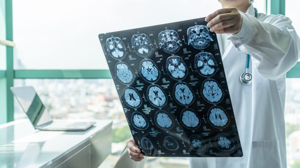
MRI of your brain will give AD researchers valuable information regarding age-related brain effects that may relate to dementia. MRI can reveal brain volume loss, brain blood flow abnormalities, and interruptions in brain function, connections or networks.
Do I have to pay for the MRI scan?
The MRI scans for individuals participating in the Cleveland Alzheimer’s Disease Research Center (CADRC) are covered by a grant from the National Institutes of Health, and there is no cost to you.
Positron Emission Tomography (PET):
What is a PET scan?
Positron Emission Tomography (PET) is an imaging method using a tiny amount of a radioactive tracer (chemical substance that interacts with cells or tissue) that emits a type of radiation that can be seen by the scanner.
PET is typically performed as a combined PET and Computed Tomography (CT) scan. The CT portion of the exam is used to improve the accuracy of the PET images and is not performed as a diagnostic-quality scan.
How long does the radiation last?
The radioactive tracer used in this research decays rapidly, with a half-life of under two hours. After 24 hours, the radiation is no longer detectable and is less than the natural background radiation level of the environment.
Why does dementia research include a PET scan?
PET allows doctors to see changes at the molecular level, often before these changes can appear in an MRI.
For Alzheimer’s disease research, an amyloid PET scan reveals the amount and location of b-amyloid protein accumulation in the brain. b-amyloid accumulation is an early marker of Alzheimer’s disease, appearing several years before any clinical symptoms might appear. However the presence of b-amyloid does not determine that one will definitely develop Alzheimer’s disease.
How do I prepare for the PET scan?
There is no special preparation or diet for the PET scan. (Other types of PET scans do have specific instructions, but the amyloid brain PET scan does not.)
Do I have to worry about an allergic reaction? I had a reaction to a CT scan in the past.
Unlike CT scans with contrast, the amount of PET tracer injected is very small. Allergic reactions from PET tracers are very rare. The CT scan associated with the PET/CT exam does not involve contrast.
How long does the brain PET/CT scan take?
The total time for the entire procedure is about 90 minutes, but most of that time is preparation for the actual scan. The exam starts with the injection of the tracer, which takes around 10 minutes. An intravenous (IV) line is set up in the forearm, and then the tracer is injected through the IV line. After tracer injection, an individual is asked to rest comfortably in a waiting area for about 40 minutes. The individual will have the opportunity to use the bathroom before the scan.
The positioning scan and CT scan each take a few seconds. The PET scan then follows for 20 minutes.
How difficult is it to have a PET scan?
The opening of a PET scanner is 28 to 31 inches in diameter, and claustrophobia is not common. There are no loud noises during the scan, besides moderate buzzing and motor sounds during the CT part of the exam.
What are the risks of PET?
The PET/CT scan involves radiation from the tracer and from the CT part of the scan. The total amount of radiation from the brain PET/CT scan is comparable to that of other diagnostic imaging studies. The insertion of the IV line involves some discomfort and possible bruising.
Do I have to pay for the PET scan?
The PET scans for individuals participating in the Cleveland Alzheimer’s Disease Research Center (CADRC) are covered by a grant from the National Institutes of Health, and there is no cost to you.