MRI has been studied as a diagnostic tool in Alzheimer’s Disease (AD) for decades, with thousands of scans collected from older individuals, both healthy and cognitively impaired, through various population studies. Ongoing advancements continue to refine MRI methods to better capture the neurodegenerative features of AD. As part of the CADRC, you can participate in two different MRI scan sessions at multiple timepoints, each designed with distinct purposes to enhance research insights.
Scan Session 1 focuses on acquiring MRI data comparable to that collected in large-scale studies, such as the Alzheimer’s Disease Neuroimaging Initiative (ADNI). This allows our data to complement and integrate with these larger datasets, fostering broader comparisons and contextual analysis.
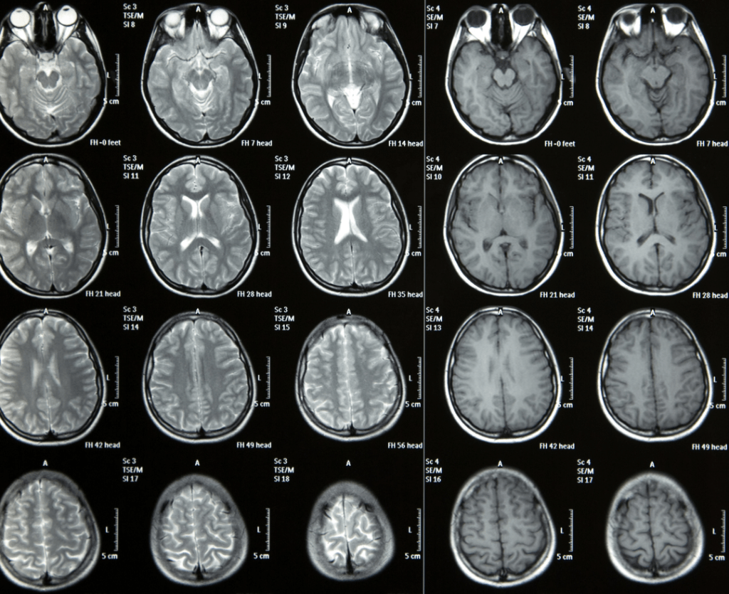
Scan Session 2 employs advanced MRI techniques recently developed in Cleveland and other research hubs. These methods provide quantitative data specific to neurodegenerative changes linked to AD and related disorders. For example:

- Myelin Imaging: Myelin, the fatty substance surrounding axons, enhances neuronal signal efficiency. Deficits in myelin are associated with neurodegeneration. This session includes MRI techniques to measure myelin content regionally in the brain.
- Blood-Brain Barrier Analysis: Subtle defects in blood-brain barrier permeability, which can affect tissue perfusion and are linked to cognitive impairment, are assessed using a novel method developed to map permeability across the brain.

In simple terms, Scan Session 1 uses well-established techniques to match data from larger studies, while Scan Session 2 explores new, cutting-edge methods to better understand the brain changes linked to AD. Both scans provide valuable information, and by completing both, you’ll help us build a more complete picture of how MRI can improve the diagnosis and understanding of AD and related conditions.